Fixation Hints
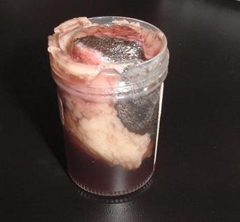
Traditionally it has been advised to use a 10:1 ratio of the volume of fixative to the volume of the sample. In practicality, we can see adequate fixation with a lower ratio, but, as seen in the above photo, samples that nearly fill the container will not have adequate fixative to prevent artifact. Fixation of large samples can present a challenge, as, regardless of the volume of fixative, the penetration of formalin is limited to a few cm.
Large samples
— in some cases, sectioning prior to fixation may be needed with samples
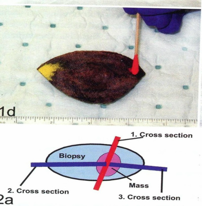
larger than a few cm in diameter. Sectioning can be done prior to fixation while retaining the integrity of the closest surgical margins. With elliptical excisions that are not too large a 1 to 2 cm thick slice section across the sample (red line in figure 2a) will
provide adequate fixation while retaining the attached closest surgical margins
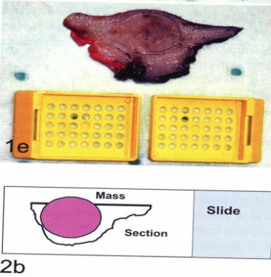
laterally and the entire deep margin. We can then produce a section that will have the closest margins of the ellipse and the deep margin, as in figure 2b. This figure illustrates the cross section through the tissue in a diagram form as well as a photo of the trimmed sample. With very large lesions, it may be necessary to choose smaller sections that include the most worrisome or closest margins. Particularly with sections of large masses, you can mark the marginal areas with special tissue marking inks or with sutures. The specimen in figure 2b has been marked partially with red ink and partially with black ink. Tissue marking is particularly helpful with smaller sections of large lesions, as shrinkage and folding may occur during fixation and orientation may be difficult. If you have bulk formalin you can keep the unsubmitted portion of a large mass on site in case additional tissue is needed for diagnosis or margins.
Figure 2a - from From: Kamstock et al. Vet Path 48:24
Figure 2b - from From: Kamstock et al. Vet Path 48:24


